 Need help to IDENTIFY
  #216446
7 y
9,215
† Cross †
Lugol’s Iodine Free S&H
J.Crow’s® Lugol’s Iodine Solution. Restore lost reserves.
Heart Worms?
Hulda Clark Cleanses
Advertisement
J.Crow’s® Lugol’s Iodine
Free S&H.Restore lost reserves.J.CROW’S®Lugol’s Iodine Solution
Fiber laden parasites:


//www.curezone.org/upload/Parasites/tn-DSCF1963_Copy

Can someone identify these parasites? (PLEASE!?) Pictures taken a few weeks ago.The one against the dark background fell out of me or my dogs. Dang! It was shooting out that white fine fiber by the truckload! That was the day I saw firsthand several parasites fall out of my forehead wound. They're the 2 pic'd together look like mini "seahorses", and they're also seen with other debris off the floor next to me the same day, pictured with the dime.
Been in this hell since last June, 2016. These pics were taken after various treatments. Last summer into fall, dogs and I used oral Ivermectin and Valbazen, each 2 times a day for about 60 days, along with Doxycycline. Doxy given to dogs with meal, yet the lab threw hers up. Any ideas of an alterntive to Doxy?
What are the mother f**, if anyone knows? Dogs had maybe still do, scabies. Older dog had crusted front "knees" for 2 years, so has the long treatment canine scabies, and possibly an underlying immune system--maybe Cushings.Don't know for sure. Property has had an onslaught of fungus issues in the plants and soils. Under the house the soil looks as if frosted with yellow powdered sugar. How to treat? Dogs had eye discharge of brown crust and red/brown watery discharge for about 3 years, same time frame of when plants and soils became fungus laden. Some creepy flies showed up in rear yard--they hang out in one place close to patio door, then attack and bite tenaciously-no fear of people. One bite a bloody hole in my hand within 15 seconds. A few weeks later I had painful section of skin behind my neck, sun on it was PAINFUL.It had thousands of eggs and larvae in it--then began the fight of my life--earlobes, cheekbone, jawline, ner chin, lips, MOUTH and gums.
Wondering why scabies seems to precede the invasion of systemic parasites? I definitely had scabies in the beginning.
Began treating our caged bird for birdmites a few months ago. Bird dropping white dandruff, is that dead bird mites? The shedding was going on too long, so I added more oral ivermectin to it's bird seed, heavy dandruff accompanied with large (for a bird) worms in it's stool. Glad I added more Iver or the worms would have gone ndetected. Going on the 3rd week of bird de-worming, dandruff almost gone. The bird mites took off into the crevices of my house.
Someone wrote "don't use the "Neem Oil Extract" Garden type. I wanted to ask why? It has warnings not for human or pet use, wash off skin immed" etc. I called the # on the bottle, cust svc said by Fed law they have to write that on all products, that it isn't harmful to people. It has 70% Neem, 30% other proprietary ingredients, some of which make it dissolve in water readily, but none are toxic. She said it any were harmful, by law it HAS to be listed separately with it's % and by name, as an active ingred. Wanted to passs that good news on, since it's only $10 for a 16 ounce bottle of concentrate.Tight on money so am grateful to discover Neem. Used it on a q-tip on some bloody sore gums, and pulled out blue fibers and globs of parasite gel-globs-eggs and young larvae. Still don't know if this invasion since July is FLY or strongyloids. Please help identify from the pics. Thanks!Advertisement
The Bio Cleanse
Complete detox kit for removing toxins, heavy metals, parasites and mucoid plaque.
- Re: Need help to IDENTIFY Cimber38
7 y
8,103
Lugol’s Iodine Free S&H
J.Crow’s® Lugol’s Iodine Solution. Restore lost reserves.
Heart Worms?
Hulda Clark Cleanses
This is a reply to # 2,353,678It looks like a fluke of some type.I would buy hot shot pest strips and hang them where the flies are. They works gewat and kill all flying and crawling insects including mites.- Re: Need help to IDENTIFY #216446
7 y
7,969
Lugol’s Iodine Free S&H
J.Crow’s® Lugol’s Iodine Solution. Restore lost reserves.
Heart Worms?
Hulda Clark Cleanses
This is a reply to # 2,353,809I get tiny flies around me indoors. I had my spray bottle and got one, it fell inches from me. It was a TINY black gnat, maybe it hatched from under the sofa or from me. I'll try to post a pic of it. - Re: Need help to IDENTIFY #216446
7 y
7,583
Lugol’s Iodine Free S&H
J.Crow’s® Lugol’s Iodine Solution. Restore lost reserves.
Heart Worms?
Hulda Clark Cleanses
This is a reply to # 2,353,809Am going to get some of those Hot Shot Pest Strips when I can. Just for outdoor use? I spray for flies inside with Malathion, zero tolerance for flies. Pest strip probably safer than Malathion. Gnats know and love me when I go outside for a few seconds. When the dogs were coming in from outside recently, one normal size fly was on the dog's fur, and intended to come in riding on the dog!! Not a normal fly!!! No fear of people.It was probably laying eggs in the fur, wish I'd bathed her right away. I wasn't using cattle topicals yet.
- Re: Need help to IDENTIFY #216446
7 y
7,969
- Re: Need help to IDENTIFY bettieblue
7 y
8,365
Lugol’s Iodine Free S&H
J.Crow’s® Lugol’s Iodine Solution. Restore lost reserves.
Heart Worms?
Hulda Clark Cleanses
This is a reply to # 2,353,678I'm sorry I can help identify what's going on but I can tell you I had similar issues involving my skin and mite looking creatures and ones similar to your images for roughly three years that eventually led me to discover my infestation had become systematic. About six months ago I started doing coffee enemas and found the same black specks along wih a plethera of other intestinal parasites. I have also struggled with my face being attacked and it's a nightmare. I'm about to post an update on my treatment. But just wanted to comment that I'd seen these creatures emerge from the sores that cover my forearms.Advertisement
J.Crow’s® Lugol’s Iodine
Free S&H.Restore lost reserves.J.CROW’S®Lugol’s Iodine Solution
- Re: Need help to IDENTIFY #216446
7 y
7,978
Lugol’s Iodine Free S&H
J.Crow’s® Lugol’s Iodine Solution. Restore lost reserves.
Heart Worms?
Hulda Clark Cleanses
This is a reply to # 2,353,818Advertisement
The Bio Cleanse
Complete detox kit for removing toxins, heavy metals, parasites and mucoid plaque.
Thanks for the replies. I hope you post your update. In the beginning my forearms were the worst, there was something (I thought a mite) in every hair follicle, plus big sores that didn't heal and had yellow plastic-like covering of whatever....looked like dried wood glue-the yellow kind-and as hard to tug off as dried woood glue. That was hell. - Re: Need help to IDENTIFY #216446
7 y
7,936
Lugol’s Iodine Free S&H
J.Crow’s® Lugol’s Iodine Solution. Restore lost reserves.
Heart Worms?
Hulda Clark Cleanses
This is a reply to # 2,353,818Was hoping some of the posters (Sharkman, Clew) may have some input. PLEASE?
Sharkman, you say you had strongyloids for several years, does any of it's larval stages look like my picture with the dark background?- Re: Need help to IDENTIFY CLEW
7 y
9,368
This is a reply to # 2,354,789Strongyloides stercoralis
1. Strongyloides stercoralis
2. Synonym Strongyloides instestinalis Anguillula stercoralis Common Name Threadworm Disease Strongyloidiasis Cochin-China Disease Geographic Distribution Cosmopolitan (lower incidence compared to hookworm) and Sporadic in temperate and cold regions which parallels Hookworm Principal Host Man Incubation Period in Man 28 days Mode of Infection Contact with the intact skin of human beings with the filariform larva; walking barefoot
3. FEMALE Parasitic Female 2.2 mm Colorless, semitransparent Filariform nematode fine striated cuticle Slender tapering anterior end, and short conical posterior end Vulva 1/3 of body length from posterior end Uteri contain 8-12 thin shelled, transparent, segmented ova Free- Living Female 1mm, smaller than parasitic Resembles typical rhabditoid free-living nematode Muscular esophageal pharynx is double-bulbed and intestine is straight Vulva 2/5 length from posterior Uteri contain a single column of thin-shelled, transparent, segmented ova
4. MALE Parasitic Male Rhabditoid in type Identical with free living male except slightly larger buccal chamber Free- Living Male 0.7mm long Tail is curved ventrad 2 equal copulatory spicules and gubernaculums No caudal end ( a protective wing-like structure)
5. LARVAE Rhabditoid Larvae Feeding stage of the parasite Open mouth, short, and stout Club-shaped anterior portion with a post median constriction and a posterior bulbous esophagus Relatively conspicuous primordium on the ventral side halfway down the midgut Buccal cavity is short and of small diameter Molt 4 times before becoming an adult Filariform Larvae Non-feeding stage Close mouth, long, delicate, and slender Has long esophagus Tail with notched or blunt or fork appearance Infective to man Can swim in water, and survive in water or soil for several threads
6. Egg or Ova Ovoid Thin shelled Transparent Partially embryonated Hatch in mucosal epithelium Strongyloides stercoralis is an ovoviviparous
7. Mode of Infection Penetration on bare skin Disease Strongyloidiasis, Cochin China diarrhea
8. Clinical Manifestations Dermatitis, swelling, itching, larva currens and mild hemorrhage at the site where the skin has been penetrated Pnuemonia-like symptoms Lofflers syndrome Tissue damage, sempsis and ulcers Hyperinfection syndrome has a mortality rate of close to 90%
9. LIFE CYCLE What are the 2 types of life cycle?
10. Specimen Feces Sputum Duodenal aspirates Gastric aspirates
11. Diagnostic Stages: S. sterocoralis eggs = Papanicolau stained smears of duodenal or gastric aspirate Filariform Larvae = Ascitic Fluid, CSF, Feces and Sputum Rhabditiod Larvae = Stools, duodenal aspirates and sputum
12. Immunologic Test Indirect hemagglutination Enzyme-linked immunosorbent assay (ELIZA) Treatment Ivermectin with albendazole (uncomplicated strongyloidiasis) Ideal method would be prevention by improved sanitation (proper disposal of feces) Practice good hygiene (washing of hand is the right manner)
http://www.slideshare.net/HazelMarieBarcela/strongyloides-stercoralis-57455693
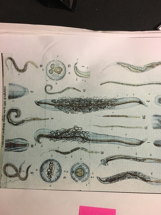
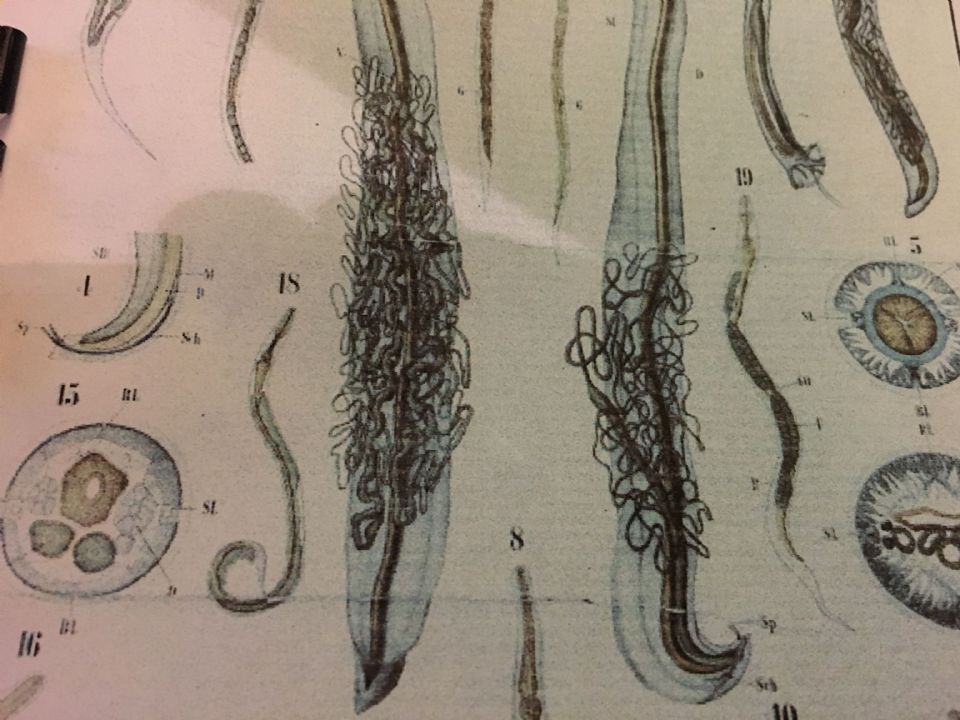
I can't say for sure if yours are strongyloides, but I think this vintage Science poster from Ernst Haeckel has some similar "hairy" looking "morg" worms:

- Re: Need help to IDENTIFY #216446
7 y
7,788
This is a reply to # 2,355,250Advertisement
J.Crow’s® Lugol’s Iodine
Free S&H.Restore lost reserves.J.CROW’S®Lugol’s Iodine Solution
Thanks Clew. I'll look at the Image Gallery for some pictures. Am having great trouble last 8 months finding on the web anywhere, good photos of parasite eggs, all larvae stages, 3rd stage would be of special interest, and adult stage.
Any place that gives a comprehensive rundown on some or many parasitic worms and mites, but esp worms, showing what it looks like in its stages from egg to adult, and where it likes to reside in the body, for example-- neck up, nose and eyes, feet and hands, just torso, etc.. If the medicine prescribed for it would be given that would be a bonus.
Advertisement
The Bio Cleanse
Complete detox kit for removing toxins, heavy metals, parasites and mucoid plaque.
- old sharkman post Kaladonia Cairns
7 y
7,630
This is a reply to # 2,355,250Great article thank you.
For when strongloides disseminate look for sharkman post from last year.
Chronic Stongyloidiasis-don't look & you wont find.
RACGP 2016
- Re: old sharkman post #216446
7 y
7,556
This is a reply to # 2,356,583I looked and couldn't find! Really. What should I put in the search box? Can you post the pictures, or tell where to find them?
- Re: old sharkman post #216446
7 y
7,435
- Re: old sharkman post #216446
7 y
7,971
This is a reply to # 2,356,583Could you kindly post the Curezone URL for Sharkman's Strongyloid article from last year? I cannot find it, thank you.
- Re: old sharkman post SHARKMAN 7 y 8,009
- Re: old sharkman post #216446
7 y
7,556
- Re: Need help to IDENTIFY #216446
7 y
7,788
- Re: Need help to IDENTIFY CLEW
7 y
9,368
- Re: Need help to IDENTIFY #216446
7 y
7,940
This is a reply to # 2,353,818Thanks Bettiblue, three years! So sorry you had to go thru that. Thanks for the coffee enema info. I do want to do this too. Seems to be what people say to use for ropeworms. which I'm suspecting as well, I think I have many different varieties of parasites. Did you get rid of them, or most of them? Did you use medications along with the enemas?
It seems an impossible battle because the eggs are all over my house. Anything left on the counter becomes infested immediately. I am using Moxidectin pour on for the dogs and myself externally, daily. The things die right in their fur that stuff works! Thanks again.Advertisement
J.Crow’s® Lugol’s Iodine
Free S&H.Restore lost reserves.J.CROW’S®Lugol’s Iodine Solution
- Re: Need help to IDENTIFY #216446
7 y
7,978
- minocycline is alternative to doxycy... sadbuttrue
7 y
7,824
This is a reply to # 2,353,678The alternative to doxy is minocycline, brand name Solodyn.
~sadbuttrue- Re: minocycline is alternative to do... #216446
7 y
7,664
This is a reply to # 2,356,249Thank you. Do you know if it's just as effective as Doxy, but with less side effects?
- Re: minocycline is alternative to do... sadbuttrue
7 y
7,803
This is a reply to # 2,356,352Advertisement
The Bio Cleanse
Complete detox kit for removing toxins, heavy metals, parasites and mucoid plaque.
According to various studies, mino is as effective or moreso than doxy. Mino is much harder to obtain tho, and is expensive.
No stomach upset as with doxy, and also no diet restrictions i.e. calcium.
Not made for animals that I know of.
If you can convince your doctor, it is prescribed long term for acne.- Re: minocycline is alternative to do... #216446
7 y
7,705
This is a reply to # 2,356,365Sadbuttrue, good to know about its advantages, my love of dairy interfered with my taking Doxy.
- doxycycline kills Wolbachia sadbuttrue
7 y
7,908
This is a reply to # 2,356,380Doxy is a very important component of a treatment regimen for certain worms. Yes, doxy kills bacteria, but why it is important is that it kills bacteria in the worm--Wolbachia--that the worm cannot survive and/or reproduce without.
If you can't get minocycline, take doxy in the middle of the night and at midday, not at meal times. Expand 22 messages, 21 to 42 of 42
  #216446
7 y
7,534
- doxycycline kills Wolbachia sadbuttrue
7 y
7,908
- Re: minocycline is alternative to do... #216446
7 y
7,705
- Re: minocycline is alternative to do... sadbuttrue
7 y
7,803
- Re: minocycline is alternative to do... #216446
7 y
7,664
- Re: Need help to IDENTIFY Cimber38
7 y
8,103
Translate This Page:


