Fungi pics
On this site they given a "warning" on these and other pics, especially if you're not the other kind of MD. I believe all of us have seen more disgusting things than these pictures. Anyway, all the pictures are good to view and I only picked a few. These fungi are all Aspergillus -- check out the the cornea scraping of the fungus.
http://www.njmoldinspection.com/human_aspergillosis.html
*More recent reports indicate that fungal sinusitis is more common than previously thought. Fulminant Aspergillosis is usually seen in immunosuppressed patients, and chronic invasive Aspergillus sinusitis, allergic Aspergillus sinusitis and Aspergilloma are seen in the non-immunocompromised individual.
Lung, Aspergillus Infection
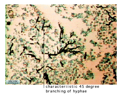
Aspergillus is an opportunistic fungus. It is an ascomycete and therefore is characterized by dimorphism (yeast forms and hyphae forms), septate hyphae, sexual spores found in an ascus and asexual spores (conidia) found on conidiophores. Aspergillus can be identified quickly by its septated hyphae and the 45 degree angle branching of its hyphae.
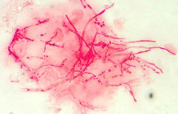
Allergic Fungal Sinusitis
Also as eosinophilic fungal rhinosinusitis, light microscopy of mucus sections. Histological sections of formalin fixed and paraffin embedded mucus, from patients suffering from allergic fungal sinusitis (eosinophilic fungal rhinosinusitis).
Kindly provided by Jens Ponikau, Mayo Clinic. Copyright Mayo Foundation 2003
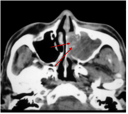
MRI Fungal Sinusitus
Calicifications in imaging. Allergic fungal sinusitis (AFS) is commonly caused by Aspergillus. Patients often have associated asthma. The MRI in addition to radiological abnormalities, thick pus or a clay-like substance was found in the sinuses. There was no allergic mucin, but dense hyphae are found. An inflammatory response in the mucosa was noted. Upon looking into the sinus, the fungus ball can vary in size from 1 mm or smaller to a size which completely occupies the sinus. It may have a greenish-black appearance.
Removal of the fungus ball is the typical treatment.
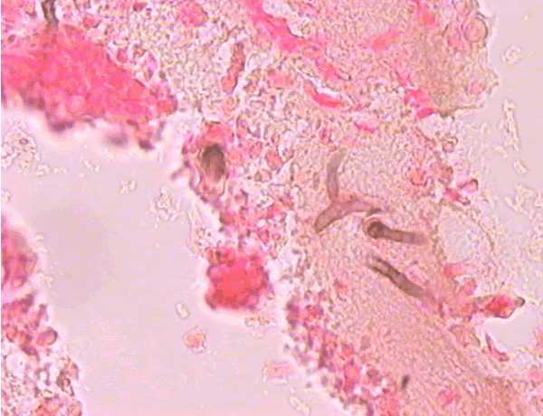
Corneal Ulcer - Gram Stain
Corneal scrapings were taken from a 67 yr old farmer presenting with a corneal ulcer of the right eye. A piece of vegetable matter was embedded in the cornea and scrapings were done. Gram stain (500x magnification) showed numerous septate hyphae. Cultures grew a small amount of A fumigatus
Kindly provided by Dr Ken Stottler (© Fungal Research Trust)
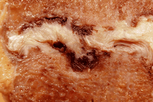
Lumbar Discitis
Macroscopic lesion of a lumbar intervertebral vertebral at autopsy, showing haemorrhagic necrosis, caused by hyphal invasion and infarction. The confirmation of genus and species was obtained by PCR on blood and vegetations; the discitis was a manifestation of disseminated aspergillosis.
Submitted by Dr R. Barnes and Dr C. Poynton, Cardiff.
 R rabbitears
17 y
12,362
R rabbitears
17 y
12,362