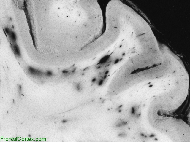
I was thinking about headaches in the frontal lobe from iodine therapy. I was wondering how the frontal lobe might be involved in behavior, and thinking about the various emotional disruptions that iodine frequently brings about. Wondering what the connection might be, and came up with these associations.
Some of the common symptoms of disorder in the frontal lobe are:
http://www.neuroskills.com/tbi/bfrontal.shtml

|
The frontal lobes are considered our emotional control center and home to our personality. There is no other part of the brain where lesions can cause such a wide variety of symptoms (Kolb & Wishaw, 1990). The frontal lobes are involved in motor function, problem solving, spontaneity, memory, language, initiation, judgement, impulse control, and social and sexua| behavior. The frontal lobes are extremely vulnerable to injury due to their location at the front of the cranium, proximity to the sphenoid wing and their large size. MRI studies have shown that the frontal area is the most common region of injury following mild to moderate traumatic brain injury (Levin et al., 1987). There are important asymmetrical differences in the frontal lobes. The left frontal lobe is involved in controlling language related movement, whereas the right frontal lobe plays a role in non-verbal abilities. Some researchers emphasize that this rule is not absolute and that with many people, both lobes are involved in nearly all behavior. Disturbance of motor function is typically characterized by loss of fine movements and strength of the arms, hands and fingers (Kuypers, 1981). Complex chains of motor movement also seem to be controlled by the frontal lobes (Leonard et al., 1988). Patients with frontal lobe damage exhibit little spontaneous facial expression, which points to the role of the frontal lobes in facial expression (Kolb & Milner, 1981). Broca's Aphasia, or difficulty in speaking, has been associated with frontal damage by Brown (1972). An interesting phenomenon of frontal lobe damage is the insignificant effect it can have on traditional IQ testing. Researchers believe that this may have to do with IQ tests typically assessing convergent rather than divergent thinking. Frontal lobe damage seems to have an impact on divergent thinking, or flexibility and problem solving ability. There is also evidence showing lingering interference with attention and memory even after good recovery from a TBI (Stuss et al., 1985). Another area often associated with frontal damage is that of "behavioral sponteneity." Kolb & Milner (1981) found that individual with frontal damage displayed fewer spontaneous facial movements, spoke fewer words (left frontal lesions) or excessively (right frontal lesions). One of the most common characteristics of frontal lobe damage is difficulty in interpreting feedback from the environment. Perseverating on a response (Milner, 1964), risk taking, and non-compliance with rules (Miller, 1985), and impaired associated learning (using external cues to help guide behavior) (Drewe, 1975) are a few examples of this type of deficit. The frontal lobes are also thought to play a part in our spatial orientation, including our body's orientation in space (Semmes et al., 1963). One of the most common effects of frontal damage can be a dramatic change in social behavior. A person's personality can undergo significant changes after an injury to the frontal lobes, especially when both lobes are involved. There are some differences in the left versus right frontal lobes in this area. Left frontal damage usually manifests as pseudodepression and right frontal damage as pseudopsychopathic (Blumer and Benson, 1975). Sexual behavior can also be effected by frontal lesions. Orbital frontal damage can introduce abnormal sexua| behavior, while dorolateral lesions may reduce sexua| interest (Walker and Blummer, 1975). Some common tests for frontal lobe function are: Wisconsin Card Sorting (response inhibition); Finger Tapping (motor skills); Token Test (language skills). Additional Material:
References: Blumer, D., & Benson, D. Personality changes with frontal and temporal lobe lesions. In D. Benson and D. Blumer, eds. Psychiatric Aspects of Neurologic Disease. New York: Grune & Stratton, 1975. Brown, J. Aphasia, Apraxia and Agnosia. Springfield, IL: Charles C. Thomas, 1972. Drewe, E. (1975). Go-no-go learning after frontal lobe lesion in humans. Cortex, 11:8-16. Kolb, B., & Milner, B. (1981). Performance of complex arm and facial movements after focal brain lesions. Neuropsychologia, 19:505-514. Kuypers, H. Anatomy of the descending pathways. In V. Brooks, ed. The Nervous System, Handbook of Physiology, vol. 2. Baltimore: Williams and Wilkins, 1981. Leonard, G., Jones, L., & Milner, B. (1988). Residual impairment in handgrip strength after unilateral frontal-lobe lesions. Neuropsychologia, 26:555-564. Levin et al. (1987). Magnetic resonance imaging and computerized tomography in relation to the neurobehavioral sequelae of mild and moderate head injuries. Journal of Neurosurgery, 66, 706-713. Miller, L. (1985). Cognitive risk taking after frontal or temporal lobectomy. I. The synthesis of fragmented visual information. Neuropsychologia, 23:359-369. Milner, B. Some effects of frontal lobectomy in man. In J. Warren and K. Akert, eds. The Frontal Granular Cortex and Behavior. New York: McGraw-Hill, 1964. Semmes, J., Weinstein, S., Ghent, L., & Teuber, H. (1963). Impaired orientation in personal and extrapersonal space. Brain, 86:747-772. Stuss, D. et al. (1985). Subtle neuropsychological deficits in patients with good recovery after closed head injury. Neurosurgery, 17, 41-47. Walker, E., & Blumer, D. The localization of sex in the brain. In K.J. Zulch, O. Creutzfeldt, and G. Galbraith, eds. Cerebral Localization, Berlin and New York: Springer-Verlag, 1975. |
I'm just thinking as well:)
http://www.gulflink.osd.mil/library/randrep/pb_paper/mr1018.2.chap10.html
Regional cerebral blood flow, that is, blood flow to different parts of the brain, may also be altered with bromism. Regional cerebral blood flow was assessed in a case of bromide psychosis using radioactive xenon (133Xe) inhalation (Berglund, Nielsen, et al., 1977). On the first exam, when the serum bromide level was 45 mEq/L (extremely high, within the potentially lethal range), the cerebral blood flow was reduced to approximately one-third of normal, with abnormal regional flow characterized by low flow in regions of the cortex, including frontal and parieto-occipital regions. Dialysis led to improvement in the clinical condition, and restoration of regional cerebral blood flow (Berglund, Nielsen, et al., 1977). Changes in regional cerebral blood flow--reflecting or perhaps influencing altered regional neuronal activity in the brain--could relate to symptoms of bromism."
...............................................................................................................................................................
Last updated on Thursday, March 26 2009 by gliageek

The steady-state distribution of bromide (Br−) in the nervous system of the rabbit was studied at various plasma concentration levels (0.5–20 mm). In addition, utilizing a ventriculo-cisternal perfusion system, the flux of Br− between CSF and blood was measured under various experimental conditions. It was observed that the steady-state distribution of Br− in brain and cerebrospinal fluid was concentration dependent and that brain served as a “sink” for the plasma and CSF. The results of the flux experiments revealed that the efflux of Br− from the perfusion fluid was some 30% greater than the influx of Br− from the blood. In addition, the movement of Br− out of the perfusate was attenuated with increasing concentrations of Br− and on death of the animal. The results suggest that, at low concentration levels, Br− is rapidly cleared from the CSF (by an active transport system) and the brain (by either an active transport system or oxidation of Br− to BrO3−), while at high concentration levels, the relative ineffectiveness of the proposed systems results in the accumulation of Br− and the establishment of a state of equilibrium between blood, brain and CSF.
Yes, perfect. So, bromine slowly comes out, the more we have, and over time, as levels lower, the speed of exit increases until a new equilibrium is established. Sounds like what Cougar has described clinically, that his bromine excretion levels have increased the longer he's been on iodine. Thanks for this. :)