Brain Cell Damage From Amino Acid Isolates by Aharleygyrl .....
A new sweetener, Neotame, has chemical properties of known toxicity to man.
Date: 9/27/2007 5:14:25 PM ( 17 y ago)
NutraSweet ~ Equal ~
"Sugar Free" ~ Neotame
Protein is nature's building blocks of life. Protein is used for producing and maintaining muscle, tissue, blood, hormones, enzymes including the body's organs, skin, and for healing processes.
Proteins are large, complex organic compounds made up of many groups of amino acids linked together. There have been twenty-two (22) amino acids identified as necessary for normal human growth and development.
The body can make fourteen (14) of these amino acids, and these are called non-essential amino acids. The other eight (8) amino acids must be received through outside sources, as in the foods we eat. These amino acids are called essential amino acids.
Proteins are broken down during the process of digestion. These proteins are broken down over time into their component amino acids or into very small groups. The amino acids are then used by the body for maintaining health.
Amino acids also play a key role in neurotransmission, solute concentration and balance (especially in areas of the brain), cellular calcium pump (gate) effectors, production and expenditure of ATP (the cell's energy stores), and are involved in overall body nerve cell conduction systems.
The amino acids that are released into the blood stream are competitive. This means that the various types of amino acids compete for attachment sites on enzymes and cell structures. It is this competition, which restricts any one type of amino acid from becoming too dominant and causing an imbalance in the normal ratio of the different circulating or cellular amino acids.
The enzymes, which are located throughout the body, including the brain and nerve cells, are responsible for ensuring that the amino acids gets to their proper end destination to be utilized by the body tissues.
Many key factors, including food additive excitotoxins and environmental poisons, play a role in nervous system degeneration. Collected evidence and accumulated non-industry funded research leaves no doubt that the powerful excitotoxin, aspartame and its breakdown products, have a central or predominant role in creating or exacerbating neurodegenerative or neurocarcinogenic diseases.
The focus of our report, is an overview of excitotoxic effects upon brain chemistry due to Aspartame's amino acid isolates.
Amino acid isolates have been artificially separated from the rest of the protein chain and are part of the Aspartame compound. Aspartame is then added to foods during the manufacturing process. Thus, these amino acids are by themselves (isolated), as single or dipeptide molecules. This is very different than the 80-300 amino acid chains that form natural proteins from dietary sources.
Some examples of genetically modified (rDNA) or manufactured amino acid isolates are glutamic acid or glutamate (i.e. monosodium glutamate or MSG), aspartic acid or aspartate, and phenylalanine, among others.
The isolates differ from dietary (with food) amino acids because dietary amino acids are absorbed from the gut. This depends upon the body's digestive action to break down the long amino chains (proteins) and then to absorb them. This means, that through the body's regulation of metabolism, the proteins are broken down slowly, and always in the nutritious mix of other amino acids in the proper enzyme-regulated proportion for use by the body.
Following digestion of normal proteins, the broken amino acid chains are slowly released into the body. Since they are in competition with one another for the enzyme sites, as earlier discussed, the body ensures that no one amino acid dominates the others. Although, it has been noted that phenylalanine is the strongest competitor for many of these enzyme sites.
Moreover, the effect of a dosed amino acid isolate cannot be used in synthesis of proteins in the same manner as dietary amino acids, because the body requires the " variety of the mix" to prepare and manufacture proteins, including the availability of the different enzymes and intermediary structures.
The excitotoxic effects of glutamic acid isolates are well studied and widely known. Some beneficial uses of amino acid isolates, such as L-lysine for use against oral herpes virus (SI) are also well known.
It is important to recognize the difference between natural, dietary amino acids, and pharmaceutically produced (including rDNA) amino acid isolates.
It is also important to recognize that the isolates of Aspartame are incorporated into a compound containing free methanol, a dangerous carcinogen and mutagen which readily breaks down into formaldehyde and formates inside the human body.
The hazards of ignoring the pharmacological nature of amino acid isolates are best illustrated by the Phenylalanine isolate that is 50% by weight of Aspartame. A can of soda pop yields about as much Phenylalanine as a large helping of beans.
NOTE: One (1) 12oz. can of diet soda contains 200mg of aspartame.
It should also be noted that the pharmaceutical isolates of amino acids in aspartame are produced from genetically modified bacteria (E.coli).
ABOUT PHENYLALANINE:
The dietary Phenylalanine from the beans would only be harmful to the person with PKU (phenylketonuria), a condition caused by one of several enzyme deficiencies. This creates/allows increased plasma levels of Phenylalanine (overload) leading to the formation of destructive neurotoxic effects.
In healthy individuals, the fact that dietary Phenylalanine is in competition with the other amino acids and is absorbed slowly over ten to twenty hours from the digestive tract, makes it helpful rather than harmful for them.
In contrast, the Phenylalanine (isolate) from the can of aspartame-laced soda pop is absorbed in about five minutes. This goes to the portal vein in the liver, with virtually no other competitive amino acids. Amino acid release from the liver is through an enzyme-linked channel. Without any competition, this Phenylalanine is released into the blood stream as an overwhelming bolus, or flood.
Even when ingested with foods, aspartame substantially increases the plasma phenylalanine (and aspartic acid) levels, due to their pharmaceutical make-up as isolates, and due to phenylalanine's strong competitive affinity for the enzyme mediators and transmitter catalysts.
Synergistic damage also results
from the absorption-metabolism sequence of methanol --> formaldehyde --> formic
acid. Methanol and formaldehyde are carcinogenic and mutagenic, and alter
mitochondrial DNA as well as nucleic DNA through the formed adducts of these
metabolic poisons. This may be a strong initiator of disease states because the
damaged DNA may not allow the cell to function properly or maintain homeostasis.
EXCITOTOXIC AMINO ACIDS' PATHWAY TO THE BRAIN
Background: THE BRAIN
The brain, on an anatomical level, is an integrated network of nerve cells, support cells (astrocytes, oligodendrocytes, et al), and is the controller for nerve-endocrine coordinating functions and its feedback network.
The brain controls the body's endocrine system through nerve transmission, which centers on the functionality of the hypothalamus and pituitary gland. This includes the nerve-endocrine coordination of the pancreas and secretions of the adrenals, thyroid, and gonads. This, in turn, acts upon the brain and pituitary and on the tissues throughout the body. This tightly controlled system produces a wide range of effects for proper functioning of the human organism.
Some hormone effects are used for development of the organism, from conception through birth. Some are long lasting. Many are a permanent element for life. Hormones can be initiated during maturity. Some hormones act later in adult life and can signify changes in brain function, or are associated with disease states or aging.
Nerve-endocrine functions can also act upon aspects of human behavior. This integrated system signals you when you are hungry, or upset. The overall health of this system affects learning, cognitive reasoning, controls your temperature, allows you to smell fresh-baked chocolate chip cookies, stimulates growth, oversees your heart rate, and is necessary for all the life functions enjoyed and needed by the individual. The Brain, in effect, is the "Chief Operating Officer" of your physical body.
__________________________________________________
There are two avenues or pathways to furnish nutrients, oxygen, and
other selective chemicals to the brain. These are via the
Blood Brain Barrier (BBB) and/or the Cerebral
Spinal Fluid (CSF).
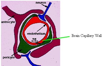
The Blood Brain Barrier is similar, in some ways, to the blood vascular network in the other parts of the body. The Blood Brain Barrier resembles normal capillaries, with a few exceptions.
The body's capillaries, outside of the brain, are more permeable (porous) to fluids, ions, and other molecular structures because there are very minute spaces between the cells making up the capillary walls.
The brain's capillary system (blood brain barrier), on the other hand, are composed of tightly packed cells or "junctions" which reduce their permeability and eliminates the bulk flow of solutes through them.
Because of the tight junctions between the blood brain barrier's capillary cells, there are two specialized ways that nutrients and other molecules can gain access to brain cell components and neurons. These pathways are: 1) lipid mediation or 2) catalyzed (active carrier) transport.
The lipid transport system is confined to the transfer of small molecules to the brain tissue, and generally are proportional to their lipid solubility.
The catalyzed transport system includes both receptor and carrier mediating enzyme processes in order to provide the brain with nutrients (glucose, amino acids, and nucleosides, etc.)
DIAGRAM #1: The Blood Brain Barrier - capillary structure and adjacent nerve cell structures.

Another function of the blood brain barrier is to isolate the brain from toxic products and certain chemicals that could disrupt the delicate balance of ions, nutrients, and neurotransmitter substances that are used by the brain's nerve cells.
When Aspartame is ingested and enters the blood stream, the three toxins of aspartame are "launched" throughout the body very rapidly.
Following consumption of aspartame-laced products, the phenylalanine flood overpowers the enzyme systems of the brain, setting off an induced PKU effect.
This induced PKU affect occurs by grossly overwhelming those enzymes required to reduce the circulating phenylalanine for use in other metabolic reactions.
This "overdose" of the competitive phenylalanine isolate (and aspartic acid) incapacitates the enzyme actions which controls several types of neurotransmitters (and their precursor amino acids) reducing dopamine and serotonin production.
The excitotoxin's effects creates secondary components which are also destructive in nature to the sensitive, surrounding neural tissues, including a breakdown by-product of phenylalanine called, diketopiperazine (DKP) which instigates tumor generation, especially that of the aggressive glioblastoma.
Further neuron insult is added due to the destruction and mutation of nucleic and mitochondrial DNA from the known carcinogenic properties of the methanol --> formaldehyde --> formic acid components.
The other avenue of delivering nutrients and other necessary molecules to the brain's cell structures is by way of the Cerebral Spinal Fluid (CSF).
There is a structure called the 'Choroid Plexus" which is a specialized arterio-venous capillary bed located within the lateral ventricles of the cerebral hemispheres that secretes the cerebral spinal fluid. (See Diagrams # 2 and # 3)
The cerebral spinal fluid is a clear, colorless liquid that circulates around the brain and spinal cord and baths the tissue with needed nutrients and other constituent molecules.
DIAGRAM #2:
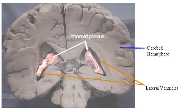
DIAGRAM #3:
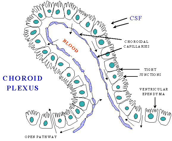
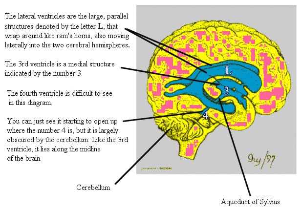
The brain is seen to contain four cavities within it. The cerebrum holds
two large Lateral Ventricles that connect at the midline. From here, the CSF
follows the InterVentricular Foramina into the Third Ventricle.
Here, at the Third Ventricle, another Choroid Plexus adds additional CSF. The CSF then passes through the Aqueduct of Sylvius, continuing into the Fourth Ventricle, located between the cerebellum and brainstem. Here, still another Choroid Plexus at the roof of the Fourth Ventricle contributes additional CSF fluid.
After leaving the Fourth Ventricle, the CSF essentially flows backward and downward around the midbrain, exiting the ventricular network below the cerebellum.
Some of the CSF passes downward into the Spinal SubArachnoid Space (circulating around the spinal cord), and a portion rises upward, through the Tentorial Notch, spreading over the hemispheres of the brain.
Re-absorption of CSF is predominantly assumed through the lymph and blood capillary network of the subarachnoid space covering the cerebral hemispheres and spinal canal.
VENTRICLE NETWORK:
DIAGRAM #4:
The nutrients supplied by the CSF are delivered by diffusion into those structures adjacent to the CSF. This leads to some specialized circumstances.
First, as the CSF diffuses nutrients (or toxins) to these adjacent structures, there becomes less and less concentrations of these molecules remaining within the CSF, as it continues along its track.
If toxins are present within the CSF, those structures first contacted are far more severely attacked by these toxins (or excitotoxins), than those bathed in CSF farther away from the ventricular system and choroid plexus.
Second, diffusion of chemicals is greatly increased by increases in hydrostatic pressures or flow rates.
Third, inflammation of the Aqueduct of Sylvius from repeated insults of neurotoxic chemicals contained within the CSF, can cause narrowing of this duct, resulting in obstruction and the onset of adult hydrocephalus.
It is in the CSF, where the phenylalanine and dicarboxylic aspartic acid diffuses, setting off a chain reaction of repeated excitatory stimuli of surrounding nerve cells and neuronal structures adjacent to the flow route of the CSF. This eventually leads to nerve cell necrosis (cell death) in these areas.
The hypothalamus sits adjacent on either side of the Third Ventricle, where there is a high diffusion rate from the CSF, leading to sustained and potentially extreme damage to this neuro-endocrine structure, one of the most vital neural systems in the body.
DIAGRAM #5:
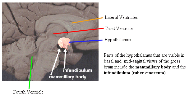
A (simplistic) sequence of events would be the following:
The transport of excitotoxins across the blood brain barrier and within the CSF causes several reactions to occur. The excitotoxins stimulate the nerves to fire excessively. The normal enzyme actions required to offset the induced, repeated firing of these neurons are negated by the phenylalanine and aspartic acid.
Furthermore, the energy system for the required enzyme reactions becomes compromised from: depleted intracellular ATP stores, the presence of formaldehyde, altered intracellular Ca+ uptake, damage to cellular mitochondria, destruction of the cellular wall, and the subsequent release of free radicals. This potentiates oxidative stress and neurodegeneration.
These toxic by-products initiate secondary damage, which increases capillary permeability, continuing to destroy the surrounding nerve and glial cells. This further impedes enzyme reactions, and promotes DNA structural defects.
Cellular death occurs over the next 1 to 12 hours. This does not include the long-term or cumulative effects of formaldehyde adducts and other metabolites. The dead cells leave behind lesions, or holes, as Dr. Olney discovered with tests he conducted. Evidence indicates that the following disease states can be clinically identified by their corresponding anatomic nerve fiber, or nerve bundle damage:
Above the Fourth Ventricle, lay the pyramids, and slightly forward are the basal ganglia. With powerful insults from excitotoxic stimulation, we develop clinical manifestations of Parkinson's disease (See Diagram # 6). This is further complicated by the depletion of the neurotransmitter, dopamine, resulting from the obliteration of enzyme sites by the flood of these excitotoxins.
Parkinson's Disease, itself, is a complex chronic brain disorder resulting primarily from progressive death of a specific group of nerve cells in a layer of a region of the substantia nigra (basal ganglia) in the midbrain.
DIAGRAM #6:
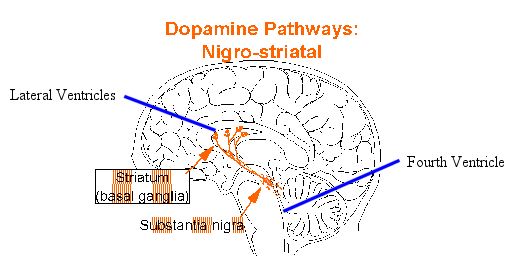
These nerve cells produce a chemical neurotransmitter called dopamine (which is inhibited by the phenylalanine/aspartic acid isolates of aspartame). The dopamine enables communication with receptors on neurons in a region of the brain called the striatum. Additional dopamine pathways run from the midbrain to the limbic area and to the cerebral cortex.
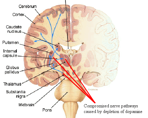
The striatum includes three structures: globus pallidus, putamen and caudate nucleus. (See Diagram 7, below)
The striatum is a part of the brain involved with regulating the intensity of coordinated muscle activity such as movement, balance and walking. Insufficient levels of dopamine from the neurons of the substantia nigra synapsing on neurons in the striatum is believed to be responsible for the primary symptoms of Parkinson's.
View of Brain structures affected by Parkinson's (and surrounding structures)
DIAGRAM #7:
As with Parkinsonian, Amyotrophic Lateral Sclerosis (ALS), commonly
called Lou Gehrig's Disease, clearly represents a connection between nerve
damage and the presence of excitotoxic amino acids.
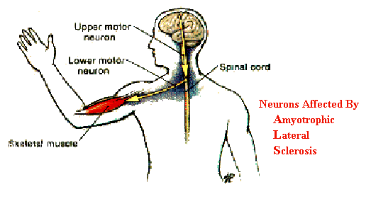
Amyotrophic Lateral sclerosis, or ALS, is a progressive, degenerative disease resulting from damage or destruction of motor neurons within the brain and spinal cord. Nerve cell destruction impairs or prevents muscle movement that corresponds to the affected neurons.
The various types of ALS include "bulbar", which affects the cranial nerves, creating complications with speech and swallowing, et al. When the damaged neurons extend from the spinal cord to muscle fibers, this is often termed motor neuron disease. There can also be various combinations of these degenerative states.
Effects of the excitatory amino acid, glutamate, have been observed in the brain and spinal cord. There is an increase of this excitotoxin within the CSF. Additionally, the damaged areas of the cerebral cortex and spinal cord fail to "uptake" this neurotransmitter substance, leaving higher amounts in the extracellular space, causing notable cellular damage due to its excitatory properties.
NOTE: Aspartate (aspartic acid) is similar to glutamate and reacts with many of the same enzyme structures.
Also noted, are pathologies related to cellular calcium (Ca+) channels, which are altered by the presence of glutamate or aspartate. Calcium changes may cause further deterioration by triggering secondary antibody effects that react to this damage. Cellular damage will cause the release of oxidizing agents, resulting in high, free radical exposure that even further damages the nerve cells. Mitochondrial damage is compounded from this surge in free radical generation.
DIAGRAM #8:
Multiple Sclerosis (MS) is a disease that affects the myelin (myelin sheath), which is the insulation or coating of some of the nerves of the brain, spinal cord, and of the periphery. Damage has also been identified that affects a part of the nerve fibers called the axons. Oligodendrocyte damage and cell loss also occurs.
Nerve cell damage takes place within the "white matter" of the brain, where the neurons have myelin sheaths, giving this part of the brain its color.
Demyelination of the central nervous system (white matter) are hallmarks of this disease. This is normally (but not always) accompanied by optic neuritis, asymmetrical muscle weakness, or fatigue.
Evidence seems to point to an immunological disorder, as a response to an inflammatory process in the brain (and/or spinal cord).
Although many scientists have not identified a definitive cause of Multiple Sclerosis, many hypothesize that MS may be of a triggered immunological origin. These authors' investigative research into known scientific endeavors, biochemical facts, and available data, leads to the following deduction. Hopefully, this will prompt further investigation into the etiology of MS by non-industry funded researchers.
IF MS is an "immunological" response, then the following need be considered:
Evidence reveals that Methanol has long been the agent most well know to cause auto-immune antibodies to "attack" the pancreas and myelin sheaths of neurons.
It is proposed that, regarding MS diagnosis' (and possibly other potential auto-immune diseases of the central nervous system), this sequence of damage from aspartame would elicit an inflammatory reaction. This leads to a generalized (and possibly defective) immune response against the neurons that have sustained damage, alteration, or exhibit other "non-familiar" DNA changes.
In some cases, the immune system itself may have been damaged by the formaldehyde's mutagenic effects or affected by brain chemistry/enzyme changes, creating a flawed system. This could cause the body to attack and catabolize its own nerve (or other) cells. Destroyed neurons will eventually be absorbed, leaving lesions or holes where they once had been.
Abstinence from aspartame appears to relieve the clinical presentation of excitotoxic-induced MS.
This immune process defect may (in part) also explain the rise in cross-chemical sensitivity syndrome.
_________________________
This is due to an incompetent blood brain barrier and neuronal (brain) damage produced by excitotoxins circulating the fetal brain areas. This is especially true for those areas adjacent to the brain's ventricular system. There is no doubt that destruction or damage of the hypothalamus and corresponding neuro-endocrine organs, leads to potential developmental complications (physical and mental).
Additionally, fetal alcohol syndrome can be
mimicked through the methanol components of aspartame, and is a direct result
from the maternal ingestion of aspartame.
Other disorders of fetal neurotoxin exposure will show up after birth, in the form of patho-physiologically induced learning and behavior disorders, attention deficit disorders, and the potential of DNA structural mutagenisis from formaldahyde concentrations, adducts, and the accompanying excitotoxic damage.
_________________________
Of Special Note:
During the production of aspartame, none of the animal studies conducted revealed the true neurotoxic nature of this poison in humans. That means that the studies were design-flawed from day one. A successful pharmaceutical firm, and seasoned intelligence or research personnel do not overlook this type of testing application by accident.
It is evidenced that, even with the relatively lower doses used during initial testing, many of these test animals still became sick or died as a result of ingesting aspartame.
Prior to the development of aspartame, it was a well known fact that phenylalanine interfered with human brain chemistry and had once been considered as a chemical warfare agent due to its neurotoxic capabilities.
Physiologically, human beings are approximately 60 times more sensitive to phenylalanine toxicity than any of the animals tested.
Furthermore, humans are 10-20 times more sensitive to methanol poisoning both as a subchronic and chronic toxin/carcinogen. On the contrary, the animals studied are more sensitive to the more common ethanol found in alcoholic beverages due to differences in enzyme concentrations of the species.
Humans are also about 8-10 times more sensitive to the affects of aspartic acid and glutamates, than the test animals being used.
__________________________________________________
NOTE:
A new sweetener known as Neotame has chemical properties of known toxicity to man.
Neotame appears to be a chemically altered "aspartame", possessing similar proportionate neurotoxic qualities.
I suspect the new name was formed to superficially distance this product from aspartame, due to the symptoms generated and because of the publicity this poison has received. Additionally, we should note that our public health agencies, as well as our regulatory policies, are in severe need of reorganization, to put it mildly...
http://www.wnho.net/aspartame_brain_damage.htm
Popularity: message viewed 3225 times
URL: http://www.curezone.org/blogs/fm.asp?i=985136
<< Return to the standard message view
Page generated on: 11/25/2024 12:05:46 PM in Dallas, Texas
www.curezone.org